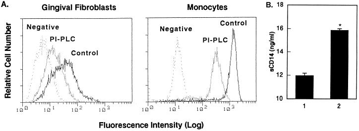
FIG. 3.
Decrease of HGF and monocyte CD14 expression by PI-PLC treatment. HGF (donor MH) from confluent cultures and MNC were suspended at 105 cells/20 μl and 106 cells/20 μl in the medium, respectively, and cultured in the presence or absence of PI-PLC (1 U/ml) for 1 h at 37°C. (A) The cells were then stained with anti-CD14 MAb (MEM-18) and analyzed by FACS analysis. The fluorescence of the monocyte population was analyzed by gating on the basis of forward/side scatter characteristics. Negative, fluorescence of negative control cells incubated with the second Ab only. (B) The amount of CD14 released in the supernatant of HGF in the absence (lane 1) or presence (lane 2) of PI-PLC was analyzed by ELISA. ∗, P < 0.05 versus lane 1. The results are representative of three different experiments.

