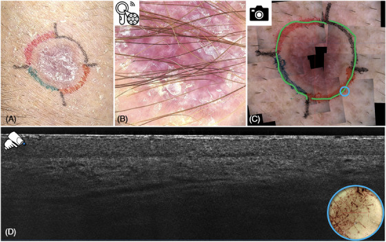
FIGURE 3.

Sclerosing basal cell carcinoma (BCC) in clinical (A) and 30× optical magnification dermoscopic view (B), as well as supplementary dermoscopic mosaic (C). The blue circle indicates the localization of the line‐field confocal optical coherence tomography (LC‐OCT) probe visualizing the corresponding vertical LC‐OCT tumor‐free image (D).
