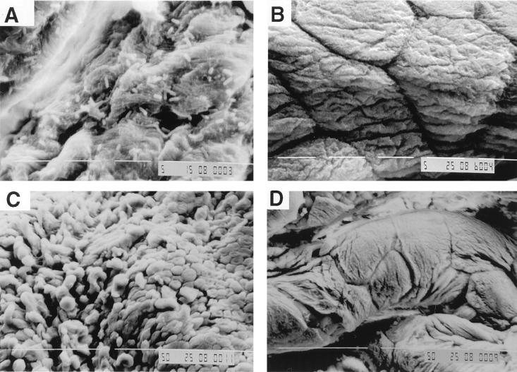
FIG. 7.
Scanning electron photomicrographs of mouse bladder mucosa after in vitro exposure to E. coli suspensions. After 1 h of exposure cells of cystitis strain f11 are observed adhering to the bladder mucosa (A) but no cells of pyelonephritis strain CFT 073 are observed (B). After 2 h of exposure to cystitis strain f11, the bladder mucosa appears to be disrupted (C), unlike mucosa exposed to pyelonephritis strain CFT 073 for 2 h (D), which appears similar to mucosa exposed to PBS (pH 7.2) for 2 h (data not shown). Magnifications, ×2,000 (A and B) and ×500 (C and D).

