. 2024 Jan 10;25(2):134–145. doi: 10.3348/kjr.2023.0769

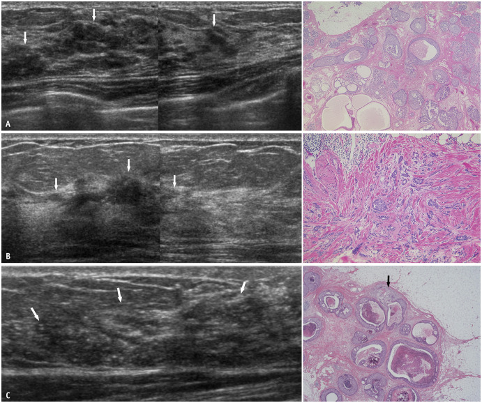
Fig. 6. Hypoechoic areas in the mammary gland. A: Intermediate-grade DCIS. US (left) shows partly patchy and partly geographic hypoechoic areas (arrows) with segmental distribution. Histopathology (right, hematoxylin and eosin stain) shows papillary-cribriform-solid growth patterns of DCIS. B: Invasive lobular carcinoma. US (left) shows a geographic hypoechoic area (arrows) with segmental distribution. The interface between adipose and glandular tissues appears to be partially interrupted. Histopathology (right, hematoxylin and eosin stain) shows small non-cohesive cells invading the stroma. C: Microinvasive carcinoma. US (left) shows an indistinct hypoechoic area (arrows) with segmental distribution. Numerous echogenic foci suggesting calcifications are present within the hypoechoic area. On histopathology (right, hematoxylin and eosin stain), most of the areas are non-invasive, comedo, solid, cribriform, papillary proliferation; however, focal areas of tumor invasion (arrow) are present. DCIS = ductal carcinoma in situ, US = ultrasound.

Fig. 6