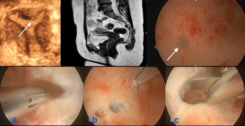
Figure 1.

Indirect and direct images in the same patient: remark the small subendometrial cyst at ultrasound in the fundal area, also seen at hysteroscopy (white arrows), absent at MRI. Remark the normal aspect from the rest of the JZ. During the operative hysteroscopic procedure 4-5 small cystic structures, not identified with indirect imaging, were opened containing the typical brownish fluid of adenomyotic cysts.
a: brownish fluid from small adenomyotic cyst; b: identification of another small cyst; c: insight view of the cyst.
