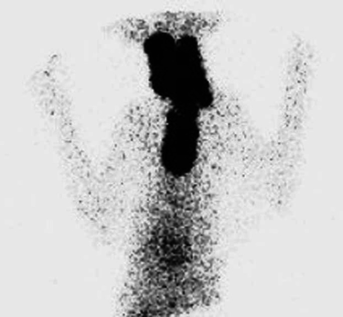
Fig 2.
Scintigraphy scan, ventral view (Table 2; case 24) showing very extensive area of IRU involving both glands in cervical area extending into cranial thorax. In such cases that involved all three areas, the areas of IRU were described as being present in the neck, thoracic inlet and thorax and a more detailed description describing the pattern of uptake was made (Table 2). This thyroid gland had a very cystic appearance on ultrasound.

