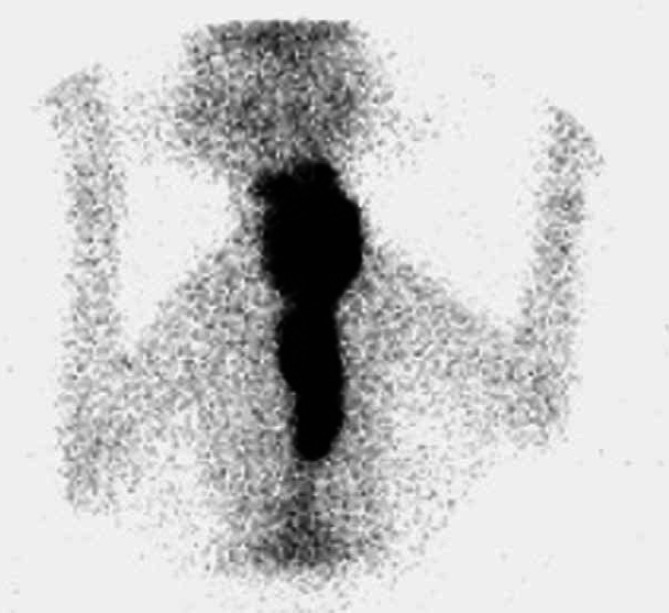
Fig 4.
Scintigraphy scan, ventral view (Table 2; case 25) showing an extensive area of IRU involving both glands in the neck and extending down the cervical region into the thorax. Biopsies of the mass in the neck confirmed adenomatous hyperplasia. The cat only partially responded to LD radioiodine treatment and went on to be well stabilised with medical management.

