
Fig 5.
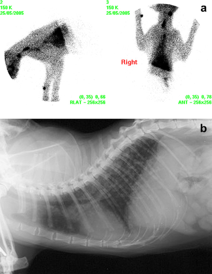
(a) Case 1 scintigraphic image – two discrete cervical regions of IRU and diffuse pulmonary uptake consistent with pulmonary metastases. (b) Case 1, thoracic radiograph (right lateral view) demonstrating diffuse bronchointerstitial pulmonary pattern.