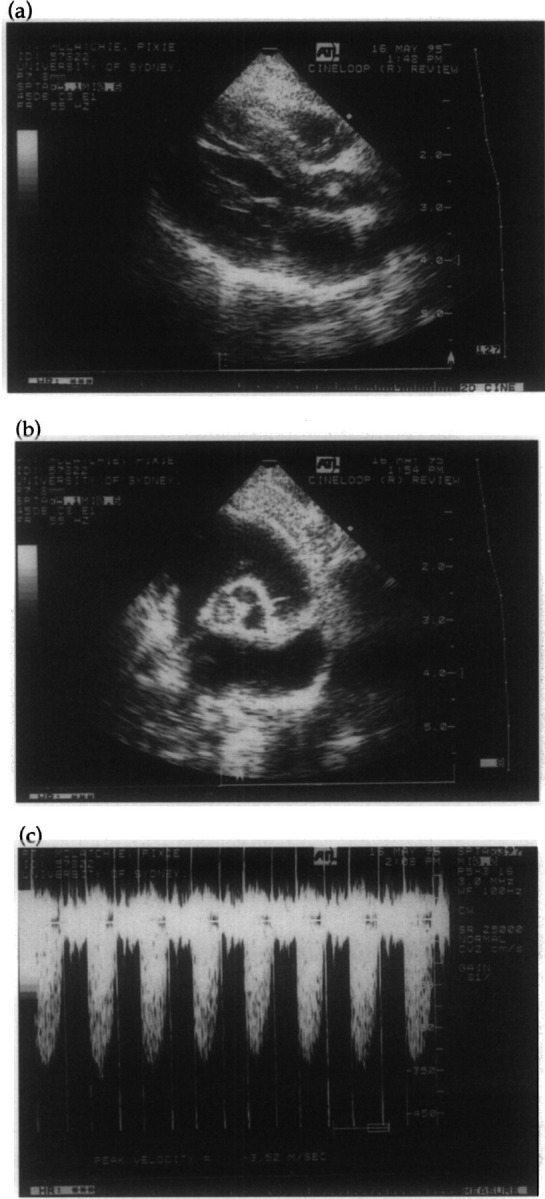
Fig 2.

Two-dimensional (a) and (b) and spectral Doppler (c) echocardiograms of Case 2 taken the day after admission to the referring veterinarian. The right parasternal long axis view (a) demonstrates thickening of the aortic valve leaflets, especially the non-coronary cusp. The parasternal short axis view (b) demonstrates thickening of the non-coronary cusp of the aortic valve. Interrogation of the aortic valve from the left caudal parasternal window using continuous wave Doppler (c) demonstrates increased peak blood flow velocity (3.5 m/s) indicative of acquired aortic stenosis.
