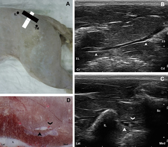
Fig 2.
(A) The anatomic landmarks for the glutea cranial approach are outlined by black dots. The position of the transducer is represented by a black rectangle for the longitudinal and a white rectangle for the transversal approaches. (B) Longitudinal US image of the ScN (arrow heads). (C) Transverse US image of the ScN (full arrow head). Another small oval structure with the same appearance corresponding to the gluteous caudalis nerve is observed (open arrow head). (D) Cross-sectional anatomical image of the ScN (full arrow head). The gluteous caudalis nerve, a branch from the truncus lumbosacralis (open arrow head) is visible. IL=os ilium; TM=trochanter major of os femoris; IS=tuber ischiadicum; Sc=os sacrum; Lat=lateral; Med=medial; Cr=cranial; Cd=caudal.

