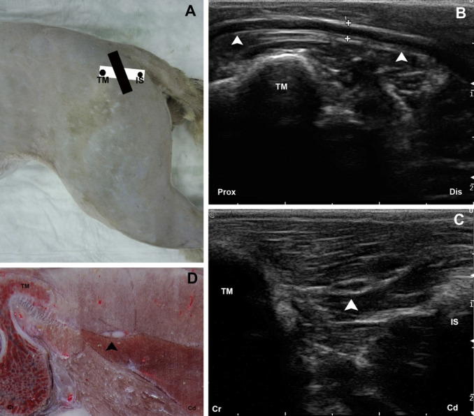
Fig 3.
(A) The anatomic landmarks for the glutea caudal approach are outlined by black dots. The position of the transducer is represented by a black rectangle for the longitudinal and a white rectangle for the transversal approaches. (B) Longitudinal US image of the ScN (arrow heads). (C) Transverse US image of the ScN (arrow head). (D) Cross-sectional anatomical image of the ScN (arrow head). TM=trochanter major of os femoris; IS=tuber ischiaticum; Prox=proximal; Dis=distal; Cr=cranial; Cd=caudal.

