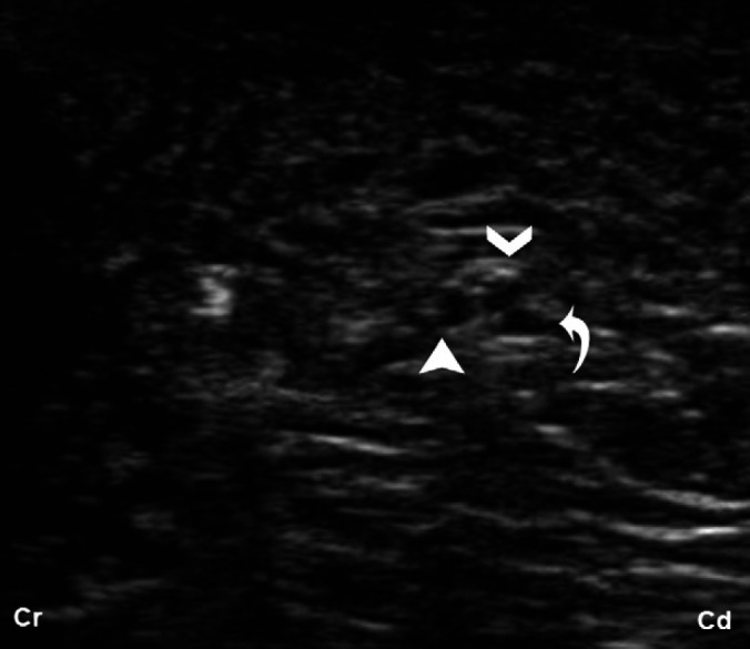
Fig 6.

Transverse US image of the ScN at midfemoral approach. At this level, three hypoechoic round structures, surrounded by a hyperechoic halo are visible, representing the peroneous communis (full arrow head), tibialis nerve (open arrow head), components of the ScN and the third structure (turned arrow) corresponding with the caudal cutaneous sural nerve branch from the tibial nerve.
