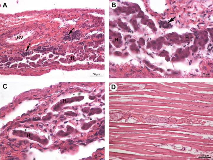
Fig 2.
Female neutered cat; light micrographs of transparent cupola of diaphragm (A–C) and intact muscular part (D). H and BV indicate the hepatocytes arranged in small groups or rows and the blood vessel. Black arrows and asterisks indicate bile ducts and sinusoidal-like spaces, respectively. HE staining.

