Abstract
In Romania, the incidence of malignant melanocytic tumors is continuously increasing. According to the World Health Organization, the incidence of melanocytic and non-melanocytic skin neoplasms has increased considerably and globally, in the last decade. We present the case of a 49-year-old patient who, over the course of 7 years, came in the Plastic Surgery Clinic of the Emergency County Hospital of Craiova for the excision of a number of 25 skin tumor formations, located on the face, cervical region, trunk and upper limbs. Treatment included complete microsurgical excision and supervision. In the end, the patient's treatment compliance decreased significantly.
Keywords: Melanoma , neoplasm , tumor formations , tumor , carcinoma „in situ” , squamous cell carcinoma
Introduction
Squamous cell carcinoma is mainly caused by exposure to the sun, so it can be diagnosed anywhere on the exposed surface of the skin.
It can also develop on skin that has been burned, damaged by chemicals or exposed to X-rays [1].
Squamous cell carcinoma is frequently found on the lips; on the surface of an old scar, but also on the epidermal area around the mouth.
About 2% to 5% of squamous cell carcinomas spread to other parts of the body, making it more aggressive than basal cell carcinoma.
On the other half, if we speak about melanomas, it had initially, an increased incidence in females.
In recent years, an increase in male cases has been observed, with 150,000 new cases being registered, mainly in the 40-60 age decade.
Healthy tissue is most frequently affected, but malignant melanoma can also appear in areas with precancerous lesions: pigmented neocellular nevi, actinic (dysplastic) nevi, giant nevi, actinic keratosis, burns.
Currently, malignant melanoma raises major problems globally, being one of the most aggressive tumor processes and compared to other malignant pathologies, it can present non-specific features that distinguish it from other types of cancerous tumors [2, 3].
Case presentation
We present the case of a 49-year-old patient, who presented himself to the Plastic Surgery Clinic of the Emergency County Hospital of Craiova, for the first time 7 years ago.
A consent from the patient has been obtained for publication of this case report.
At the age of 43, the patient presented himself to the clinic for multiple tumor formations on the face, anterior cervical region and anterior chest.
At this moment, the patient had a history of tumor formations on the face and trunk since 2016.
Upon admission to the clinic, the excision of six tumor formations was performed, where the histopathological examination established the diagnosis of carcinoma "in situ", accompanied by actinic keratosis l at the level of: nasal pyramid, right cheek, anterior chest, right supraclavicular fossa.
At the deltoid level, two formations were excised, which from an histopatologycal point of view, proved to be different.
An "in situ" carcinoma type lesion and a well-differentiated squamous carcinoma type (G1) lesion were identified at this level.
All lesions were excised within oncological safety limits.
One month later, the patient returns to the clinic, for multiple skin tumor formations located on the face.
Tumor lesions are excised from the left eyebrow, frontal region, temporal region, genial region and from the upper lip.
The tumor formation at the level of the left eyebrow arch proved to be a well-differentiated squamous carcinoma [4, 5, 6].
The tumor formation at the right temporal level proved to be a carcinoma "in situ" with Bowenoid type actinic keratosis.
All lesions were excised within oncological safety limits.
Post-operative evolution was favorable.
The patient complied with the instructions of the attending physician and no more de novo lesions were identified for a period of 4 years.
During this period, the patient was kept under specialized supervision.
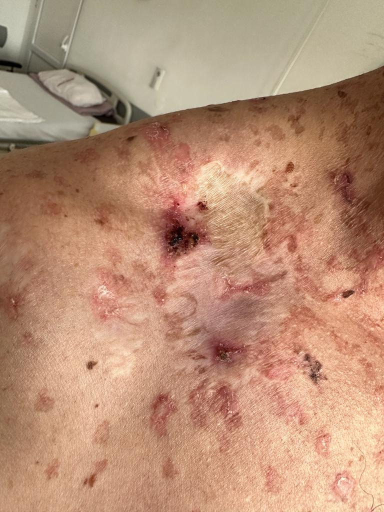
At the age of 47, after a free interval of 4 years, the patient returns to the clinic for the presence of "de novo" tumor formations located at the level of the left temporo-parietal region, right supraclavicular fossa (Figure 1), right hand dorsal front, posterior cervical region, left deltoid region, right lower eyelid region, totaling six new tumor formations.
Figure 1.

Supraclavicular fossa tumor
The tumor formations at the level of the left temporal region, left palpebral region, left supraclavicular fossa, dorsal face of the right hand was identified from an anatomo-pathological point of view as squamous carcinoma type tumors with the deep limit of surgical resection not invaded by the tumor.
The deltoid and posterior cervical tumors were identified as carcinoma "in situ" during the histopathological examination.
All lesions were excised within oncological safety limits.
The post-operative evolution was favorable.
A new free interval followed, for a period of 2 years. During this period, the patient did not return to regular check-ups.
At the age of 49, the patient returns to the clinic, due to the presence of six new tumor formations, with large sizes between 2.5-3.5cm, for which excision is performed within oncological safety limits.
Until this moment, most of the tumor formations have been identified as squamous type carcinomas, most of them being "in situ" type, and accompanied by Bowenoid actinic keratosis.
All lesions were excised within the limit of oncological safety.
From the attached images, you can see the appearance of the surrounding skin and the post-operative scars, which indicate the "de novo" nature of the lesions but also the presence of multiple pigment spots-Bowenoid actinic keratosis lesions[6].
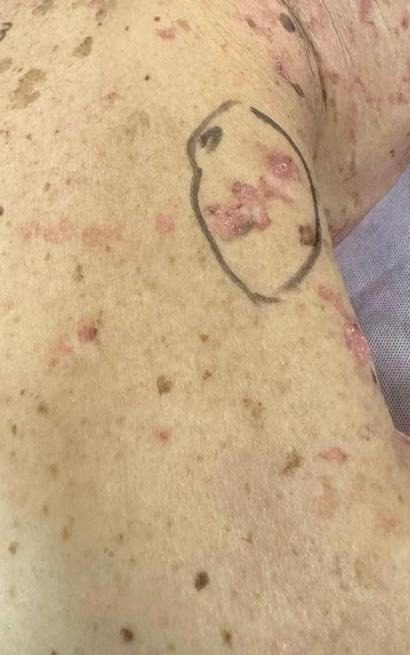
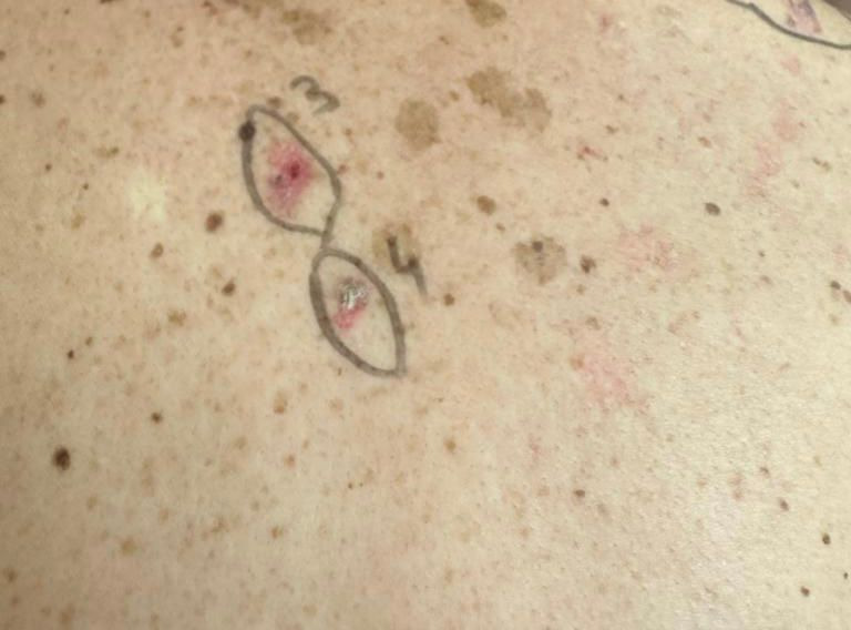
Currently, the histopathological results have highlighted five "in situ" squamous cell carcinoma tumor formations (Figure 2) and one "in situ" melanoma tumor formation-tumor number 3-(Figure 3), [7].
Figure 2.

Squamous carcinoma „in situ”.
Figure 3.

Melanoma „in situ” tumor-no 3
All tumor formations were accompanied by areas with increased melanocytic activity [8].
Discussions
The particular nature of the case refers to the multitude of tumor formations excised over a period of 7 years, but also to the appearance of the skin on the face, neck, anterior and posterior chest, where multiple lesions had been found, like bowenoid actinic keratosis lesions and keratoacanthomas tumors.
The patient's history of prolonged sun exposure highlights the need for awareness about sun protection and regular skin checks.
Public health campaigns emphasize the significance of sunscreens and protective clothing, yet there remains a substantial knowledge gap in the general population.
In recent years, the patient compliance with the treatment has dropped considerably.
The importance of consistent medical follow-up in such cases cannot be overstated.
Regular check-ups can aid in early detection and management of new lesions, reducing the risk of progression to malignancy.
Non-compliance not only jeopardizes the patient's health but also poses a significant challenge for healthcare providers striving for optimal outcomes.
In the period 2021-2023, he refused to return to periodic checks.
Such non-compliance increases the risk of unnoticed disease progression.
Currently, after the last surgical intervention, he expressed his desire to give up the treatment.
This decision may stem from fear, misinformation, or fatigue from repeated medical interventions.
Building a strong patient-doctor relationship, rooted in trust and understanding, can mitigate such challenges.
Considering the pre-existing lesions on the skin, the evolution of the patient without treatment cannot be favorable.
It's crucial for healthcare professionals to provide adequate counseling and education to such patients.
Actinic keratosis is a precancerous lesion of the skin that develops after long-term sun exposure.
So far, different series have reported variable rates of transformation into non-melanocytic cancers of the skin [9].
This is a low-grade skin tumor that typically presents as a rapidly growing, 1 to 2cm dome-shaped lesion with a central keratinous plug.
It is characterized by an initial phase of rapid growth, followed by a period of variable tumor stability.
The progression rate to squamous cell carcinoma varies between 12 and 20%.
Early intervention and consistent monitoring are key to preventing further complications.
An interdisciplinary approach involving dermatologists, oncologists, and patient counselors can provide a comprehensive care plan tailored to individual needs.
Conclusions
This case underscores the critical significance of providing comprehensive counseling and thorough education to patients diagnosed with keratoacanthoma.
Such tumors are particularly noteworthy because they undergo a phase of swift expansion, then enter a period where the tumor may remain stable for an unpredictable duration.
The unpredictable nature of its progression emphasizes the urgency of timely intervention.
Alarmingly, if these tumors are neglected and left without appropriate medical intervention, they carry the risk of advancing to squamous cell carcinoma.
Given these complexities, proactive patient engagement and continuous monitoring become paramount in ensuring optimal health outcomes and reducing potential complications.
Conflict of interests
None to declare.
References
- 1.Higgins WH, Kachiu CL, Galan A, Leffell DJ. Part I. Epidemiology, screening, and clinical features. J Am Acad Dermatol. 2015;73(2):181–190. doi: 10.1016/j.jaad.2015.04.014. [DOI] [PubMed] [Google Scholar]
- 2. Ciurea ME , Ciurea AM , Schenker M . In: Chirurgie plastica . vol I . Ciurea ME, et al., editors. București : Editura Medicală ; 2022 . Melanomul ; pp. 281 – 301 . [Google Scholar]
- 3.Marinescu A, Stepan AE, Margaritescu C, Marinescu AM, Zavoi RE, Simionescu CE. CD 44 Immunoexpression in the progression of actinic keratosis and cutaneous squamous carcinoma. Current Curr Health Sci J. 2017;43(3):241–245. doi: 10.12865/CHSJ.43.03.10. [DOI] [PMC free article] [PubMed] [Google Scholar]
- 4.Gibbons M, Ernst A, Patel A, Armbrecht E, Behshad R. Keratoacanthomas. A review of excised specimens. J Am Acad Dermatol. 2019;80(6):1794–1796. doi: 10.1016/j.jaad.2019.02.011. [DOI] [PubMed] [Google Scholar]
- 5.Jankowska-Konsur A, Kopeć-Pytlarz K, Woźniak Z, Hryncewicz-Gwóźdź A, Maj J. Multiple disseminated keratoacanthoma-like nodules: a rare form of distant metastases to the skin. Postepy Dermatol Alergol. 2018;35(5):535–537. doi: 10.5114/ada.2018.77245. [DOI] [PMC free article] [PubMed] [Google Scholar]
- 6. Bastian CB , Lazar A . In: McKee’s Pathology of the skin with clinical correlations. vol I. 4th ed . Colonje E, Brenn T, Lazar A, McKee P, et al., editors. Edinburg : Saunders Elsevier ; 2012 . MelanomA ; pp. 1221 – 1267 . [Google Scholar]
- 7.Poswar FO, Fraga CA, Farias LC, Feltenberger JD, Cruz VP, Santos SH, Silveira CM, de Paula, Guimarães AL. Immunohistochemical analysis of TIMP-3 and MMP-9 in actinic keratosis, squamous cell carcinoma of the skin, and basal cell carcinoma. Pathol Res Pract. 2013;209(11):705–709. doi: 10.1016/j.prp.2013.08.002. [DOI] [PubMed] [Google Scholar]
- 8.Schmitt JV, Miot HA. Actinic keratosis: a clinical and epidemiological revision. An Bras Dermatol. 2012;87(3):425–434. doi: 10.1590/s0365-05962012000300012. [DOI] [PubMed] [Google Scholar]
- 9.Marks R, Rennie G, Selwood TS. Malignant transformation of solar keratosis to squamous cell carcinoma. Lancet. 1988;1(8589):795–797. doi: 10.1016/s0140-6736(88)91658-3. [DOI] [PubMed] [Google Scholar]


