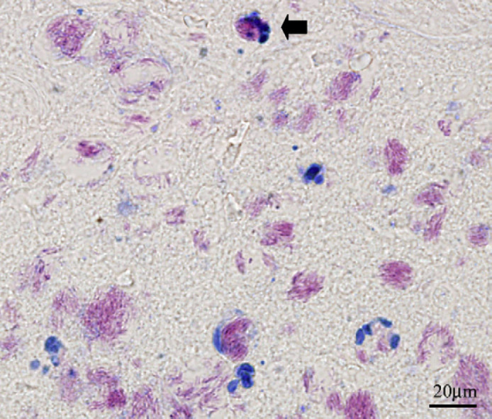
Fig 3.

ZN stained section of the lesion from a cat with a localised M ulcerans infection. Note the acid-fast bacilli (which stain pink) intracellularly within macrophages (one is labelled with an arrow). Many of the extracellular collections are of a size and shape that indicate they are derived from phagosomes originally present in cells that have undergone lysis.
