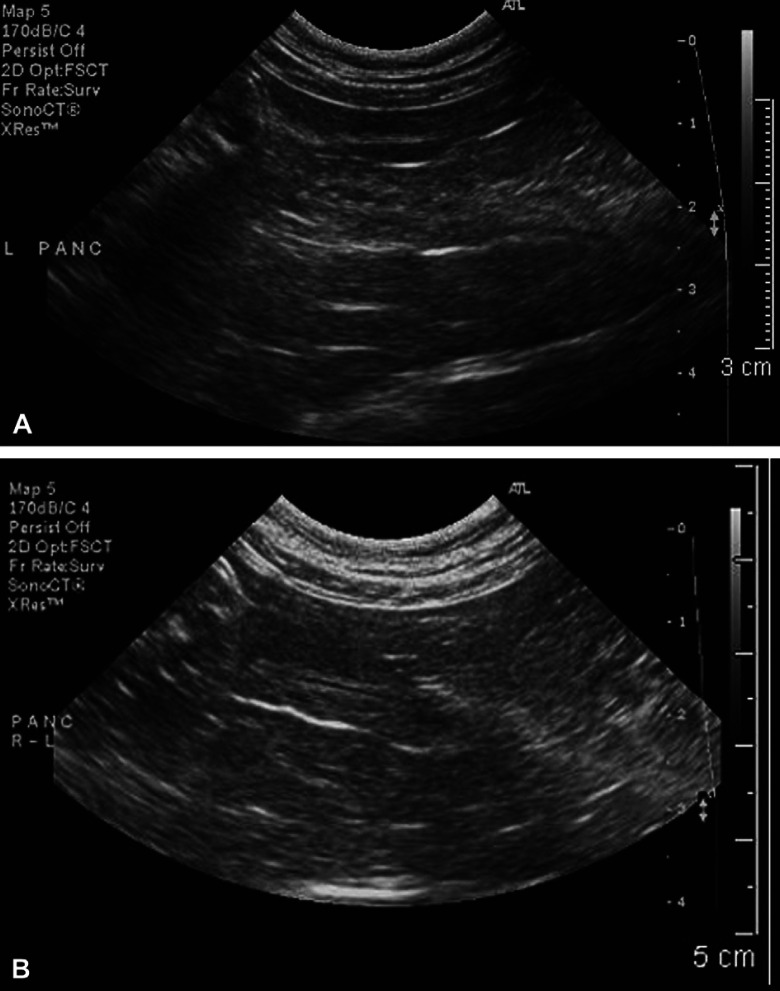
Fig 2.
Long-axis sonographic view of the left limb of the pancreas in the E procyonis infected cat. (A) At time of presentation. Note the mildly enlarged pancreas (1.0 cm thick) with its irregular contour. The pancreatic duct has thickened walls, an irregular, ‘beaded’, mildly distended lumen, and hypoechoic lumen contents. (B) Eighteen weeks after treatment with praziquantel, pyrantel, and febantel. The pancreas has a smooth contour and is reduced in size (0.8 cm thick). The pancreatic duct is now of normal size and architecture.

