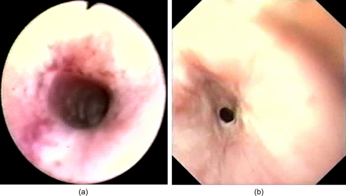
Fig 2.
(a) Oesophagoscopy prior to development of an oesophageal stricture (Case 3). Circumferential, patchy, erythema, erosion and ulceration of the oesophageal mucosa was observed 3 cm distal to the proximal oesophageal sphincter. (b) Oesophagoscopy 4 weeks later (Case 3). A benign oesophageal stricture with a lumen of 3 to 4 mm was detected 3 cm distal to the proximal oesophageal sphincter.

