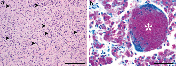
Fig 1.
(a) Typical histopathology of a cat with a mycobacterial infection. Note the densely packed polygonal to fusiform macrophages associated with a sparse neutrophilic infiltrate (arrow heads) in the lymph node of a M microti infected cat (H&E, scale bar = 100 μm). (b) Unusual histopathology of a cat with a mycobacterial infection. Note the multinucleated cell (asterisk) and very large numbers of fuchsin coloured acid-fast bacteria (AFB) in the multinucleated cell and the surrounding macrophages in the lymph node of a M avium infected cat (ZN, scale bar = 50 μ).

