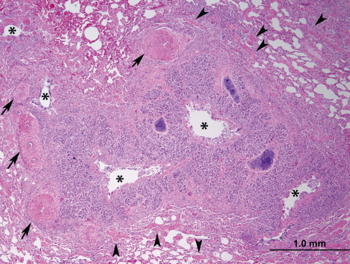
Fig 5.

Histologic section of lung. Surrounding multiple cross sections of a secondary bronchus are severely hyperplastic peribronchial glands which narrow the lumen (asterisks). Smooth muscle in the surrounding parenchyma is moderately to severely hypertrophied and hyperplastic (arrowheads). The pulmonary arteries in this case were also tortuous, evidenced by increased numbers of cross sections; the tunica media of pulmonary arteries is also moderately hyperplasic and hypertrophied (arrows).
