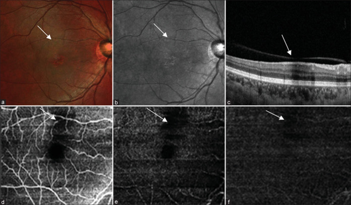
Figure 2.

(a) Multicolor composite image of the left eye shows a hyporeflective area at the posterior pole. (b) IR reflectance shows an area of hyporeflectance. (c) Structural OCT shows the area of shadowing corresponding to the area of hyporeflectance. (d-f) OCT angiography shows an area of flow void in the superficial plexus, choroidal complex, and avascular zone
