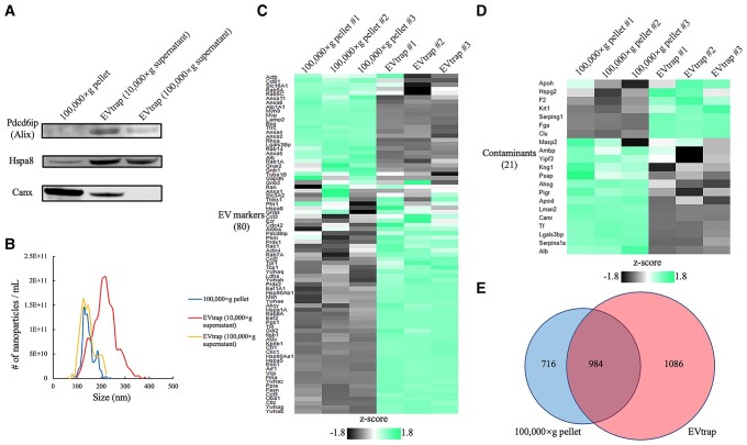
Fig. 2.
Comparison between ultracentrifugation (100,000 × g) and EVtrap for liver tissue EV isolation. Particles were isolated from 0.2 mL supernatant after 10,000 × g centrifugation on tissue homogenate. A) Western blotting data showing programed cell death six-interacting protein (Pdcd6ip, also known as Alix) and heat shock protein family A member 8 (Hspa8) as EV markers, as well as Calnexin (Canx) as contaminant protein (negative control). B) Nanoparticle analysis was performed by TRPS on 100,000 × g pellets or eluates from EVtrap beads. Relative abundances of representative C) EV markers and D) contaminant proteins by LC–MS/MS analysis with the z-score color indicated, respectively. 0.5 μg resulting peptides from each method were loaded and technical triplicates were employed. E) The numbers of identified EV proteins by ultracentrifugation and EVtrap isolation methods. Selected proteins were identified in at least two out of three technical replicates.

