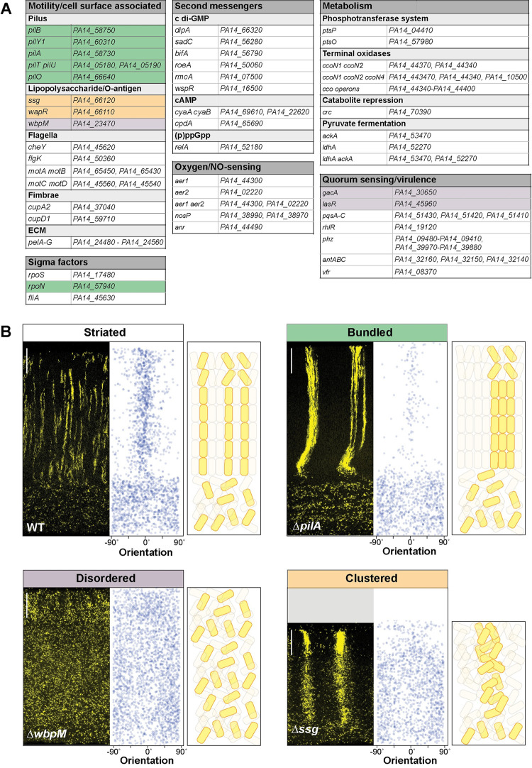
Fig 3. Specific global regulators and cell surface components are required for WT cell patterning in colony biofilms.
(A) List of genes mutated and then screened for altered cellular arrangement across depth in biofilms. Those showing altered cellular arrangement are shaded and colors correspond to the phenotype categories shown in (B). (B) Fluorescence micrographs of thin sections from WT and indicated mutant biofilms grown on 1% tryptone + 1% agar for 3 days. Biofilm inocula contained 2.5% cells that constitutively express mScarlet. mScarlet fluorescence is colored yellow. Quantification of orientation across depth is shown for each image, and cartoons of cellular arrangement are shown for each phenotype category. Images shown are representative of at least 2 independent experiments. Scale bars are 25 μm. The data underlying this figure can be found in S1 Data.

