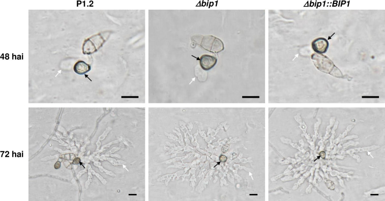
Fig 4. Appressorium-mediated penetration of Δbip1 into cellophane membranes.
Conidia of wild-type (P1.2), Δbip1 mutant and Δbip1 complemented strains were deposited on cellophane membrane, and appressoria (black arrows) and pseudo-infection hyphae growing into the membrane (white arrows) were visualized by differential interference contrast microscopy at 48 hai and 72 hai. Bar = 10μm.

