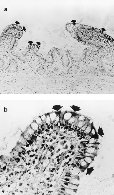
FIG. 5.
Expression of histone H1 in human terminal ileal mucosa. Immunohistochemical analysis was performed with an anti-histone H1 monoclonal antibody, but a nuclear stain was not used. (a) Histone H1 immunoreactivity was seen in the cytoplasm of epithelial cells, at the tip and on the lateral aspect of villi (arrows), as well as in nuclei. (b) Villus (from the left margin of panel a) at a high magnification. Arrows indicate immunoreactivity. Approximate magnifications: a, ×224; b, ×389.

