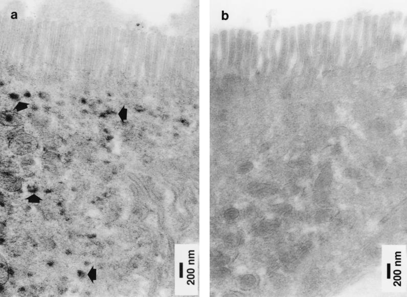
FIG. 6.
Immunoelectron microscopy of a section of normal human small intestinal mucosa labelled with an anti-human histone H1 monoclonal antibody by the peroxidase technique. Two epithelial cells near the villus tip are shown. (a) In one, immunoreactive histone H1 (arrows) was present in the cytoplasm below the microvilli. (b) The cytoplasm of an adjacent epithelial cell did not contain immunoreactive histone H1.

