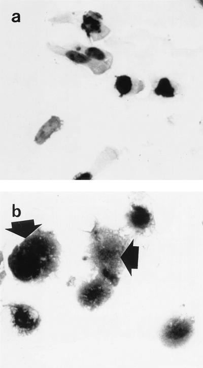
FIG. 7.
Expression of immunoreactive histone H1 in detached villus epithelial cells of human terminal ileal mucosa. Cytospin preparations of epithelial cells were made following detachment with EDTA (and subsequent washing with PBS). Photomicrographs of the same cytospin preparation were taken. Strong histone H1 immunoreactivity was seen in nuclei. Strong histone H1 immunoreactivity was seen in the cytoplasm (arrows) of some epithelial cells (b). Magnification, ×590.

