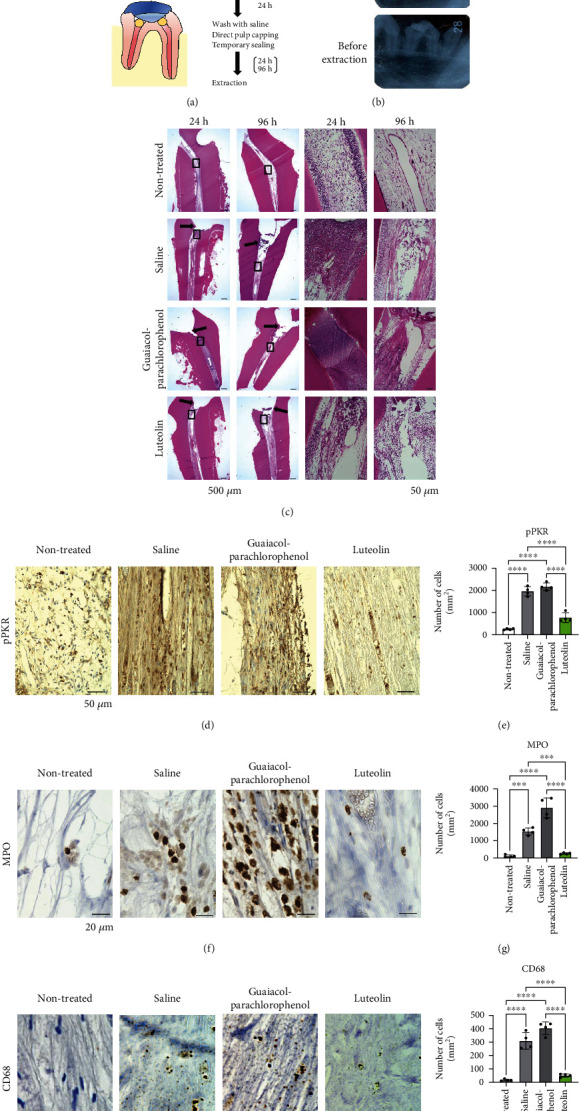
Figure 5.

Luteolin suppresses the onset of pulpitis in dog model of experimental pulpitis. (a) Schematic illustration representing dog experimental pulpitis model. After pulpotomy of the upper and lower premolars for 24 h, the root canal orifices were washed with saline and treated with luteolin- or guaiacol-parachlorophenol-soaked sterile cotton ball. After sealing the cavities for 24 or 96 h, the test teeth were extracted. (b) Dental X-ray images were captured before pulp amputation, pulp capping, and tooth extraction. (c) Hematoxylin and eosin (H&E) staining of pulp tissues was performed at 24 and 96 h after pulp capping. The arrows indicate the amputated site. Black boxes indicate magnified views. Scale bar = 500 μm and 50 μm for low (left) and high magnification, respectively. (d) Immunohistochemical staining of phosphorylated-PKR (pPKR) in pulp tissues. (e) The number of pPKR-positive cells in pulp tissue. Cell counts are indicated as positive cells per square millimeter. (f) Immunohistochemical staining of myeloperoxidase (MPO) in pulp tissues. (g) The number of MPO-positive cells in pulp tissue. Cell counts are indicated as positive cells per square millimeter. (h) Immunohistochemical staining of CD68 in pulp tissues. (i) The number of CD68-positive cells in pulp tissue. Cell counts are indicated as positive cells per square millimeter. Error bars represent the mean ± SD, n = 4. ns: not significant; ∗∗∗p < 0.001, ∗∗∗∗p < 0.0001. The statistical significance of differences between groups was determined using one-way ANOVA, followed by correction for multiple comparisons using Tukey's post hoc test.
