Figure 2.

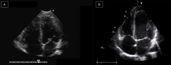
A normal patient. On the left, it’s shown an apical four-chambers view obtained with HHD (A). On the right (B), the same patient evaluated with SED.

A normal patient. On the left, it’s shown an apical four-chambers view obtained with HHD (A). On the right (B), the same patient evaluated with SED.