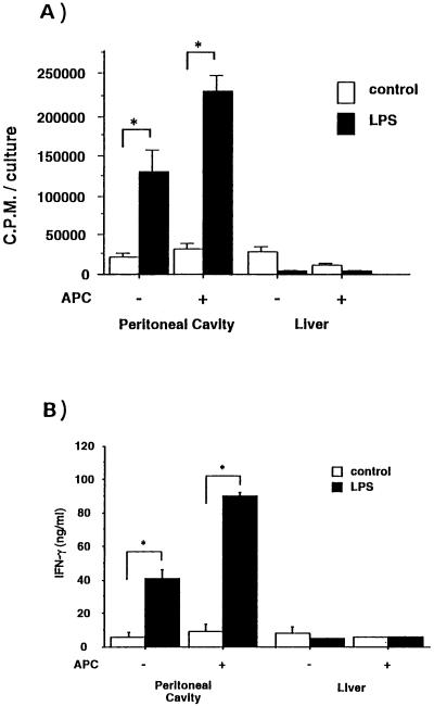
FIG. 4.
Proliferative response and cytokine production of γδ T cells from the peritoneal cavities or livers of C3H/HeN mice in the presence of LPS. (A) Purified populations of γδ cells were incubated (5 × 10⁴/well) in anti-TCR-γδ MAb-coated 96-well plates for 48 h in the presence or absence of MMC-treated spleen cells (3 × 10⁴/ml), with or without 10 μg of LPS per ml. During the last 8 h of incubation, 1.0 μCi of [3H]thymidine per well was added. The cells were then harvested, and the amount of [3H]thymidine incorporated was determined by scintillation counting. The data are representative of those from two separate experiments and are expressed as the means of triplicates ± standard deviations. Asterisks indicate significant differences from the values for the control (P < 0.05). (B) Purified γδ T cells (5 × 10⁴ cells) were cultured similarly in the presence or absence of MMC-treated spleen cells with or without LPS for 24 h at 37°C, and the culture supernatant was collected. The cytokine activity in the culture supernatant was tested for the presence of IFN-γ by ELISA. The data are representative of two separate experiments and are expressed as the means of triplicates ± standard deviations. Asterisks indicate significant differences from the values for the control (P < 0.05).

