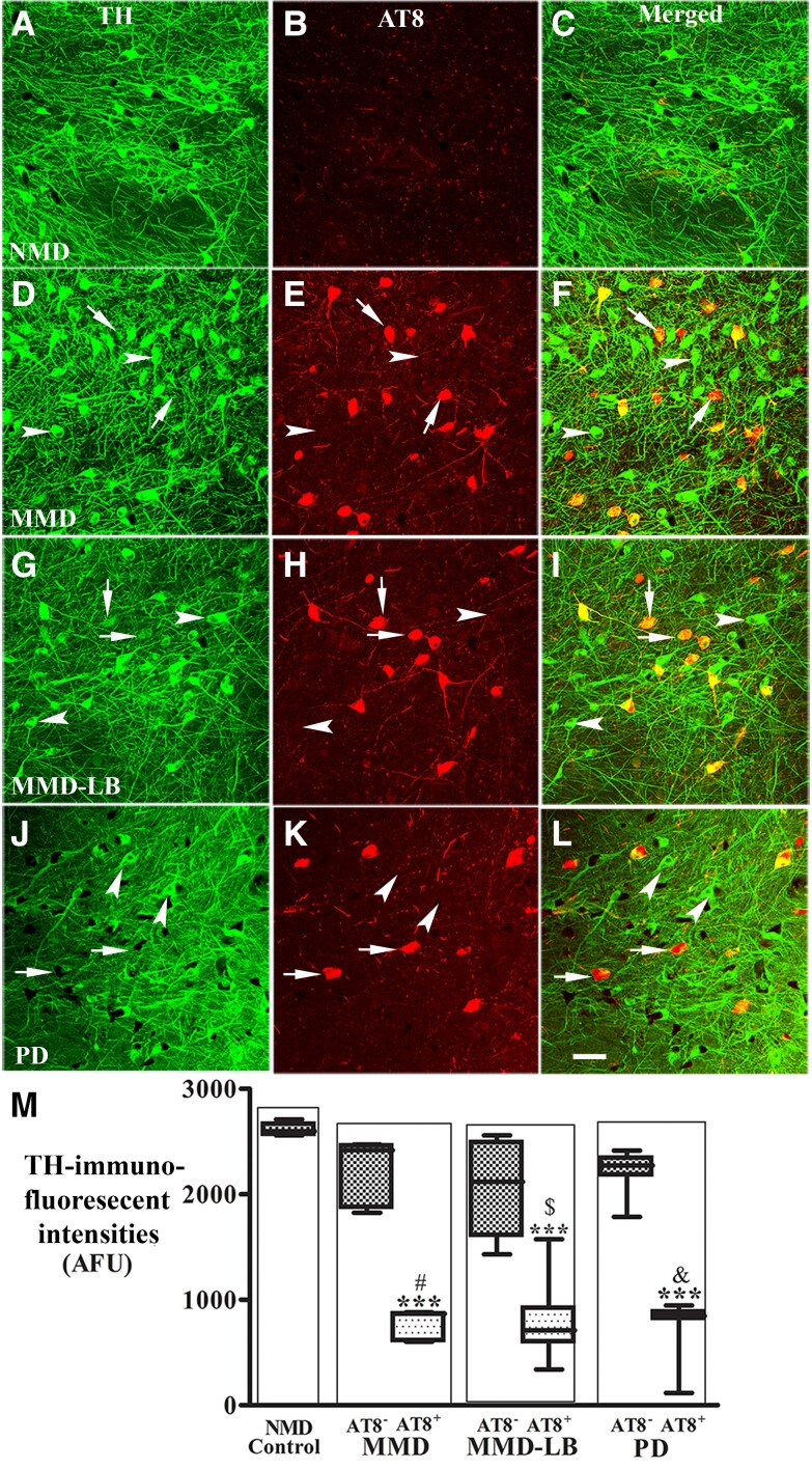
Figure 6.
Reduction of tyrosine hydroxylase levels in nigral neurons with AT8-immunorepressive aggregates. Confocal microscopic images of substantia nigra from no motor deficit (NMD; A–C), minimal motor deficits (MMD; D–F), minimal motor deficits with nigral Lewy body (MMD-LB; G–I) and Parkinson’s disease (PD; J–L) illustrated immunostaining for tyrosine hydroxylase (TH; green; A, D, G and J), AT8 (red; B, E, H and K) and co-localization of TH and AT8 (merged; C, F, I and L). Note that TH immunofluorescent intensity was extensively reduced in the neurons with tau aggregates (arrows; D–L) but not in the neurons without tau aggregates (arrowheads; D–L). Scale bar in L = 100 µm (applies to all panels). Measurements of immunofluorescent intensities (M) further revealed that TH expression was significantly reduced in the neurons with tau aggregates (AT8+) but not in the neurons without tau aggregate (AT8−). ***P < 0.001 related to NMD control, #P < 0.05 related to AT8 immunonegative neurons in MMD, $P < 0.05 related to AT8 immunonegative neurons in MMD-LB and &P < 0.05 related to AT8 immunonegative neurons in PD groups. Data: mean ± standard deviation. AFU = arbitrary fluorescence units.

