Fig. 3.

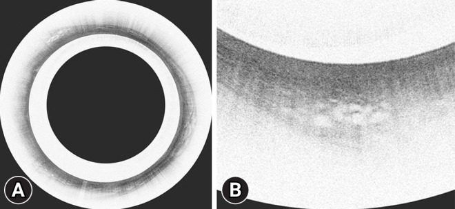
Volumetric laser endomicroscopy (VLE) images showing a classic VLE image with Barrett’s esophagus dysplasia (A) and magnified image of A showing the dysplastic area (B).

Volumetric laser endomicroscopy (VLE) images showing a classic VLE image with Barrett’s esophagus dysplasia (A) and magnified image of A showing the dysplastic area (B).