
Fig. 3.
Volumetric laser endomicroscopy (VLE) images showing a classic VLE image with Barrett’s esophagus dysplasia (A) and magnified image of A showing the dysplastic area (B).
Official websites use .gov
A
.gov website belongs to an official
government organization in the United States.
Secure .gov websites use HTTPS
A lock (
) or https:// means you've safely
connected to the .gov website. Share sensitive
information only on official, secure websites.
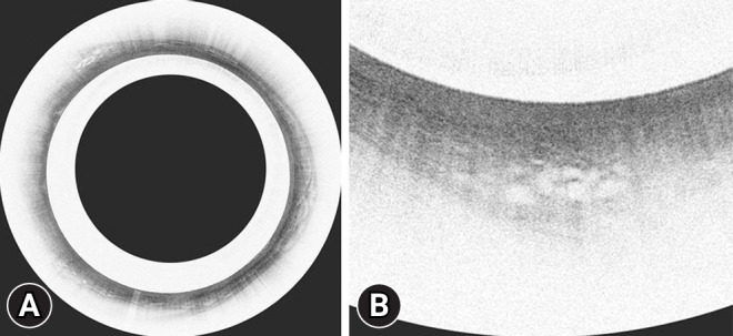
Volumetric laser endomicroscopy (VLE) images showing a classic VLE image with Barrett’s esophagus dysplasia (A) and magnified image of A showing the dysplastic area (B).