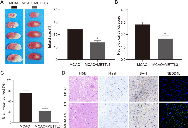
Fig. 7.
The protective effects of METTL3 overexpression in the MCAO model. A. TTC staining was used to detect the ischemic area of mice after MCAO treatment, n = 6. B. Neurological function scores of mice in each treatment group, n = 6. C. Brain water content of mice in each treatment group, n = 6. D. HE, Nissl, immunohistochemistry, and immunofluorescence were used to detect the neuronal damage and inflammatory response in the brain tissue of the two groups. * vs MCAO group P < 0.05

