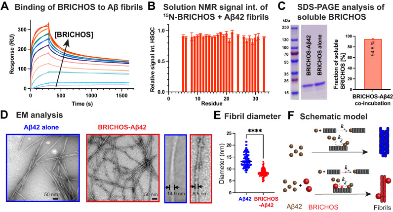
Fig. 1. Binding of BRICHOS to Aβ42 fibrils.
A SPR measurements revealing a dissociation constant of BRICHOS to Aβ42 fibrils of 12.9 ± 0.2 nM. B Solution NMR 1H-15N HSQC experiments of 15N-labeled BRICHOS showing an intensity decrease to 90 ± 8 % upon addition of Aβ42 fibrils at a 1:1 molar ratio (related to monomeric Aβ42). The error bars reflect the signal-to-noise level of one measurement (n = 1). C SDS-PAGE analysis of soluble BRICHOS after co-incubation with Aβ42 shows that the large proportion of BRICHOS is still soluble. The uncropped SDS-PAGE gel is shown in Supplementary Fig. 1. The experiment was repeated three times with qualitatively similar results. D EM images exhibiting thinner fibrils in the BRICHOS-Aβ42 sample compared to mature Aβ42 fibrils. E Fibril diameter showing a reduction of a factor of around two in the presence of BRICHOS. n = 100 independent measurements are shown where the line corresponds to the mean. An unpaired two-tailed T-test was applied, where four asterisks (****) refer to p < 0.0001. Source data are provided as Source Data file. F Schematic overview about BRICHOS-modulated Aβ42 fibril formation, where BRICHOS predominately inhibits secondary nucleation processes (k2) in addition to fibril-end elongation (k+) and favors the generation of thinner fibrils.

