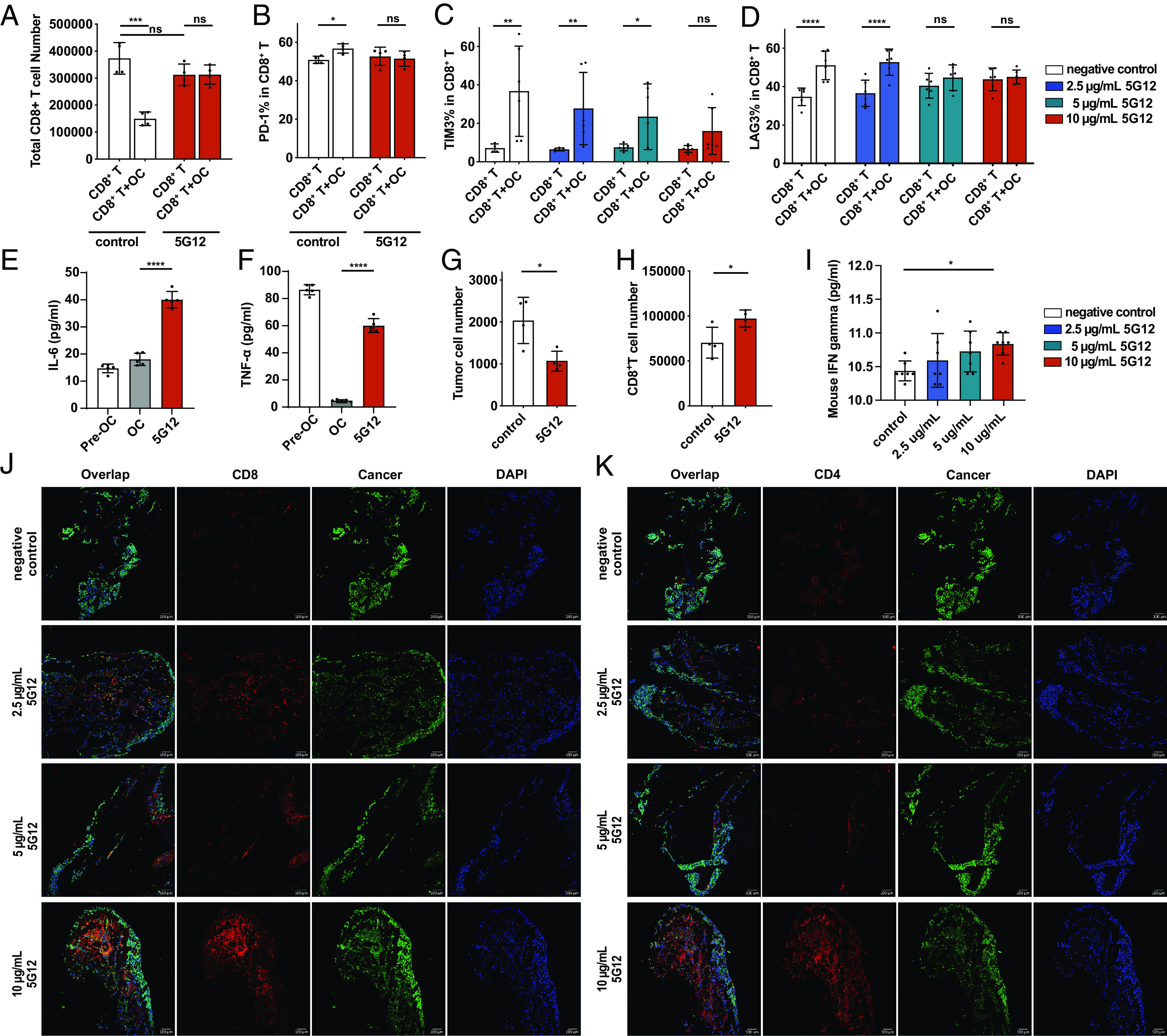
Fig. 3.

5G12 prevents osteoclast-mediated inhibition of T cells. (A) Flow cytometric quantification of CD8+ T cells after co-cultured with osteoclasts with or without 5G12 treatment, n = 4 independent experiments. The P value was calculated by the paired t-test. (B) Flow cytometric quantification of the percentage of PD-1+ CD-8+ T cells in total CD8+ T cells after co-cultured with osteoclasts with or without 5G12 treatment, n = 4 independent experiments. The P value was calculated by the paired t-test. (C and D) Flow cytometric quantification of the percentage of TIM3+ and LAG3+ CD-8+ T cells in total CD8+ T cells after co-cultured with osteoclasts with or without 5G12 treatment, respectively, n = 6 independent experiments. The P value was calculated by two-way ANOVA. (E and F) ELISAs of cytokine release of pre-osteoclasts, osteoclasts, and osteoclasts treated with 5G12, n = 6 independent experiments. The P value was calculated by the paired t-test. (G) tumor cell were counted in T cells, osteoclasts, and tumor cells co-culture with or without 5G12 treatment. (H) cytotoxic CD8+ T cells count after co-culturing T cells, osteoclasts, and tumor cells with or without 5G12 treatment. The P value was calculated by the paired t-test. (I) Quantification of mouse IFN-γ in BICA culture medium at day 7. The P value was calculated by ordinary one-way ANOVA. (J) Representative immunofluorescence staining of BICA. CD8+ T cell (red), cancer (green), and nuclei (blue). (Scale bar, 100 μm.) (K) Representative immunofluorescence staining of BICA. CD4+ T cell (red), cancer (green), and nuclei (blue). P > 0.05 [not significant (n.s.)], *P < 0.05, **P < 0.01, ***P < 0.001, and ****P < 0.0001.
