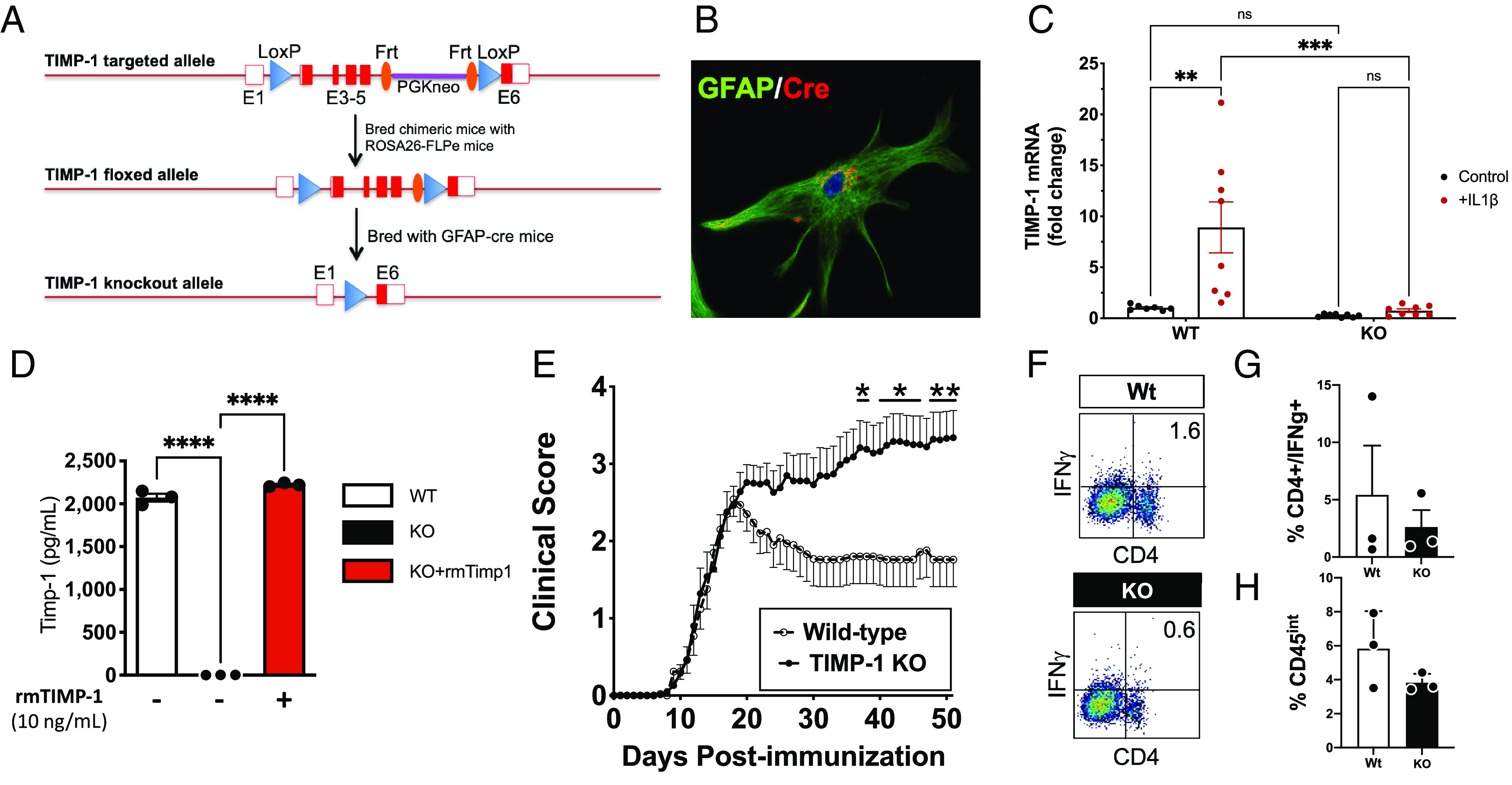
Fig. 1.

TIMP-1fl/fl knockout mice develop a severe EAE phenotype that is immunologically independent. (A) Schematic of design of development of the flox-targeted sites that enabled cell and tissue-specific deletion of TIMP-1 in GFAP-Cre-TIMP-1fl/fl mice. (B) Immunocytochemical validation of CRE expression in astrocytes from GFAP-Cre:TIMP-1fl/fl mice, and (C) physiological validation of TIMP-1 mRNA expression in primary astrocytes and absence of TIMP-1 mRNA expression in astrocyte cultures from GFAP-Cre:TIMP-1fl/fl mice (n = 8 per treatment; ANOVA; ****P < 0.0001; where *P < 0.01; ***P < 0.002). (D) Validation of absent TIMP-1 protein expression in primary astrocyte cultures from GFAP-Cre:Timp1fl/f mice (n = 3 biological replicates). (E) Clinical EAE in MOG35-55-treated C57Bl/6 WT and GFAP-Cre:TIMP-1fl/fl mice over 52 d time course (n = 13 to 17/treatment and genotype; 2-way ANOVA, P < 0.0001, where post-hoc multiple-comparisons identified significance at *P < 0.05 and **P < 0.01, as indicated). (F and G) Flow cytometry analysis of overall CD4+/IFNγ+ T cells from spinal cords of Wt and GFAP-Cre:TIMP-1fl/fl mice at peak clinical EAE illness (day 18; n = 3 per genotype and treatment), including (H) analysis of CD4+/IFNγ+ T cells (t test, unpaired t test, P < 0.20).
