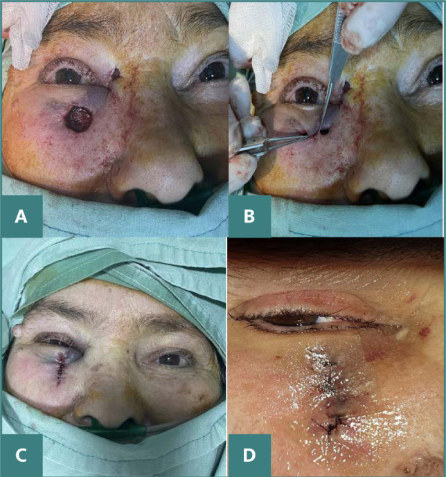
Figure 2.

Intraoperative images
A. The defect that remained following the complete excision of the keratotictumor. B. Manipulating the edge of the wound to facilitate closure of the defect, which was expanded by an additional 3 mm at the 12 o'clock position. C. Closing the defect with an advancement flap. D. Postoperative images at 10 days demonstrating satisfactory esthetic and functional results.
