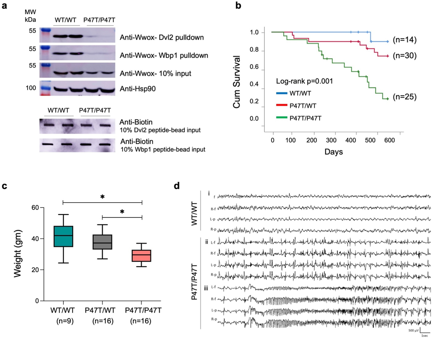
Fig. 1. |.

p.Pro47Thr mutation is sufficient to abrogate Wwox affinity for PPxY motifs. (a) Western blots showing the amount of Wwox pulled down from WwoxWT/WT (n = 2) and WwoxP47T/P47T (n = 2) CB tissue lysates using biotinylated Dvl2 (HPYSPQPPPYHELSSY) (top panel) and Wbp1 (SGSGGTPPPPYTVG) (second panel) peptides. Third panel shows Wwox expression using 10% input in WwoxWT/WT and WwoxP47T/P47T CB samples. HSP90 was used as control to demonstrate equal amount of protein used for pull-down experiment. Two lowermost panels show slot-blots of 10% biotin labeled peptide-bead input to show incubation with equal amounts of peptides. (b-d) WwoxP47T/P47T mice display reduced overall survival, hyperactive cortical spike discharges, and generalized spontaneous seizures. (b) Kaplan-Meier comparative survival in days of a cohort of WwoxWT/WT wildtype (blue line, n = 14), WwoxP47T/WT heterozygous (red line, n = 30), and WwoxP47T/P47T homozygous (green line, n = 25) mice. The WwoxP47T/P47T mice show significantly lower survival than the WwoxWT/WT and WwoxP47T/WT mice, p-value= 0.001, Log-rank (Mantel-Cox) analysis (c) Box and Whisker plot showing total body weight in grams of WwoxWT/WT (n = 9), WwoxP47T/WT (n = 16), and WwoxP47T/P47T (n = 16) mice. Box extends from 25th to 75th percentile, with horizontal line at median (50th percentile), whiskers represent bottom and top 25% and extend down to the smallest and up to the largest value, *p-value < 0.005, One-Way ANOVA (Tukey’s post-hoc test). (d) Video-EEG recording profiles showing seizure activity in representative WwoxWT/WT and WwoxP47T/P47T mice from a group of n = 3 mice/group. i. No abnormal epileptiform activity was detected in EEG from adult WwoxWT/WT littermates, ii. abnormal bilateral interictal spike discharges were frequent in WwoxP47T/P47T mutant mice, iii. generalized seizure discharge in the same mouse as ii. shows abrupt spike discharge followed by onset of fast rhythmic, and then arrhythmic patterns of cortical seizure. Traces: L-f: left frontal, R-f: right frontal, L-p: left parietal, R-p: right parietal.
