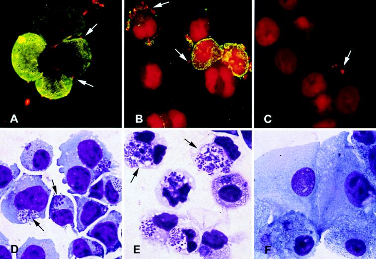
FIG. 1.
Photomicrographs of untreated and differentiated HL-60 cells incubated with the agent of HGE. (A to C) Immunofluorescence with rhodamine labeling of HGE organisms and fluorescein labeling of CD15s 15 min after HGE inoculation (magnification, ×3,300). (D to F) Giemsa stain 48 h after inoculation with HGE (magnification, ×3,300). (A) HGE binding to uninduced HL-60 cells which express high levels of CD15s; (B) increased binding of HGE to granulocytic DMSO-treated cells, particularly to those cells which most express CD15s; (C) reduced bacterial binding to TPA-treated monocytic cells which are devoid of CD15s expression; (D) infection of control uninduced HL-60 cells; (E) increased susceptibility to infection of DMSO-treated cells, as evidenced by larger and more abundant morulae; (F) absence of bacterial replication in TPA-treated cells.

