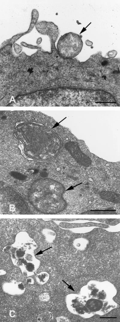
FIG. 2.
Transmission electron micrographs of untreated and differentiated HL-60 cells incubated with the agent of HGE. These micrographs were derived from the same experiment and were chosen to illustrate the altered interactions between HL-60 cells and the HGE agent resulting from differentiation. (A) Untreated HL-60 cells exhibit binding, but no internalization, of ehrlichiae 4 h after inoculation. (B) DMSO-treated granulocytic cells more rapidly internalize ehrlichiae, shown 20 min postinoculation. (C) TPA-treated monocytoid cells destroy internalized ehrlichiae, shown 2 h following inoculation. Scale bars = 0.5 μm.

