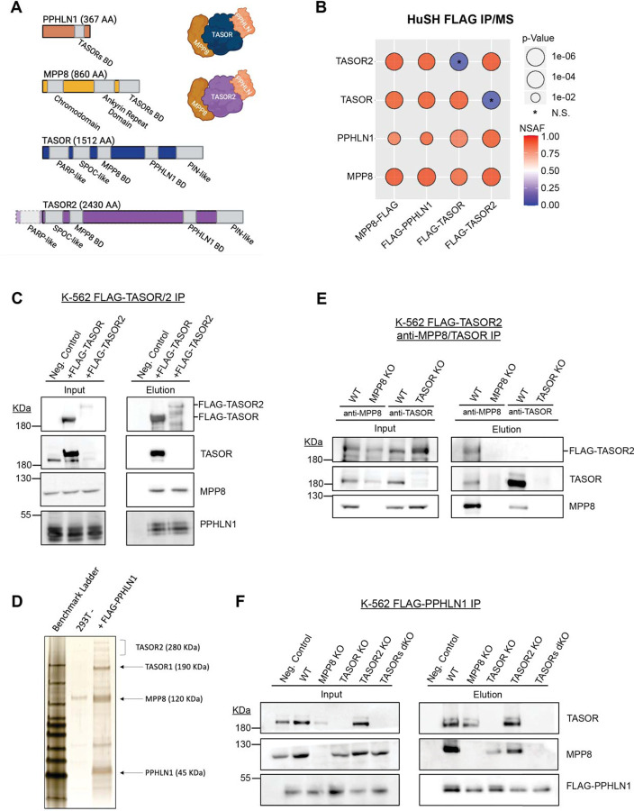
Figure 1: Identification of a second HuSH complex.
A) Molecular representation of each HuSH complex member and TASOR2 and each known domain. The Binding Domain (BD) is highlighted for reference.
B) Immunoprecipitation coupled with mass spectrometry (IP-MS) conducted in HEK293T cells expressing FLAG transgenes (x-axis) revealed the association of TASOR2 with PPHLN1 and MPP8 (y-axis). Normalized Spectral Abundance Factor (NSAF) was calculated and normalized to the length of prey targets, with comparison to the wild-type (WT) HEK293T IP negative control. IP-MS experiments were performed in bio-duplicates.
C) Immunoblots of FLAG immunoprecipitation in K-562 cell lines expressing no transgene (WT -), FLAG-TASOR, or FLAG-TASOR2, showcasing the specific interactions in the HuSH complex.
D) Silver stain of FLAG-PPHLN1 ammonium sulfate nuclear extract immunoprecipitation in 293F cells, providing visual evidence of the involvement of PPHLN1 in the newly discovered HuSH complex.
E) Immunoblots demonstrating MPP8 and TASOR immunoprecipitation in parental wild-type cells, MPP8 knockouts (KOs), or TASOR KOs expressing FLAG-TASOR2, elucidating the interactions in the context of genetic perturbations.
F) Immunoblots of FLAG immunoprecipitation performed in stable genome-edited HuSH knockout K-562 cells, expressing no transgene (Negative Control) or FLAG-PPHLN1, confirming the specificity of the interactions and the stability of the HuSH complex in a knockout background.

