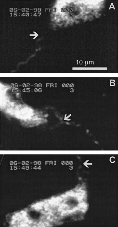
FIG. 1.
Still pictures of a video recording were captured by using a custom-made, Windows-based, frame grabber program in a PC. The pictures were further processed for printing by using Adobe Photoshop 4.0 and Corel DRAW 7. They represent a video recording, in which the attachment of the spirochete and the early events of phagocytosis can be seen. (A) A borrelia spirochete approaches a neutrophil. The arrow indicates the neutrophil-approaching end of the spirochete (time, 15:40:47). (B) The spirochete has attached to the neutrophil. The neutrophil is activated, as indicated by its elongated form. The arrow indicates the site of the attachment (time, 15:45:06). (C) The neutrophil has started to form a tubelike protrusion along the spirochete. The arrow indicates the distal end of the protrusion (time, 15:49:44).

