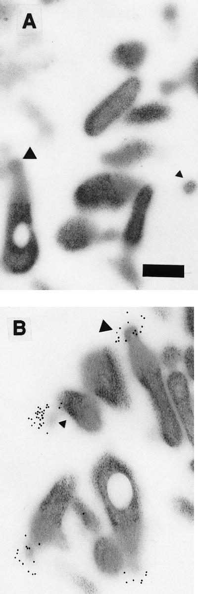
FIG. 5.
Electron micrographs showing double-sided immunogold labeling of M. gallisepticum MGC2 cytadhesin on sectioned M. gallisepticum cells. (A) Sections treated with rabbit preinoculation serum prior to incubation with 10-nm-diameter colloidal-gold-labeled goat anti-rabbit antiserum. (B) Sections reacted with rabbit anti-MGC2 antiserum. Secondary antibodies containing gold particles are distributed on the bleb organelle. The bleb regions in more longitudinal sections are visible (large triangles), while blebs in different planes of cross section are marked (small triangles). Transmission electron microscopy at ×20,000 on a Zeiss transmission electron microscope. Bar, 250 nm.

