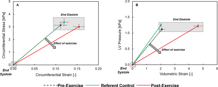
Fig 1.
(A) LV regional circumferential stress and strain was measured by speckle tracking echocardiography (STE) in which values were calculated at the onset of diastole (end-systole) and at end diastole. A rightward and downward shift in this relation, indicative of a reduction in myocardial stiffness, occurred following the exercise protocol. (B) LV volumetric strain and pressure during diastole was computed and this relation, which is indicative of LV chamber stiffness, fell downward and to the right following the exercise protocol. Values shown are composite mean values and SEM. Summary values for LV regional myocardial stiffness and chamber stiffness are shown in Table 1.

