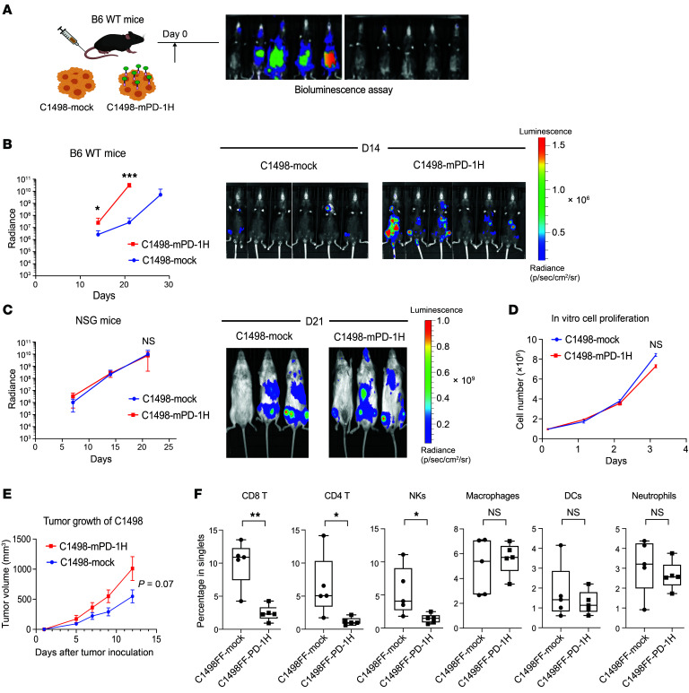
Figure 2. AML surface PD-1H inhibits T cell infiltration, leading to immune evasion.
(A) Syngeneic mouse leukemia model using tail-vein injection with myeloid leukemia cells (C1498). Mouse leukemia cells expressing PD-1H (C1498FF–PD-1H) or cells not expressing PD-1H (C1498FF-mock) were transplanted into B6 mice and assessed for in vivo leukemia proliferation using bioluminescence. (B) In vivo proliferation of C1498FF-mock versus C1498FF–PD-1H cells in B6 WT mice (n = 7). Radiance indicates the mean value per group and error bars represent SEM. P value determined by Student’s t test at each time point. *P <0.05; ***P < 0.001. These experiments were repeated 3 times. Repeated measures were determined by ANOVA with 2 factors (P > 0.05, no difference among experiments). (C) In vivo proliferation of C1498FF-mock versus C1498FF–PD-1H cells in NSG mice (n = 3) (representative images on day 21 on the right side). Radiance indicates the mean value per group, and error bars represent SEM. P value determined by Student’s t test at each time point. Repeated measures were determined by ANOVA with 2 factors (P > 0.05, no difference among experiments). (D) In vitro growth of C1498FF–PD-1H tumors compared with C1498FF-mock tumors. Statistical analysis was done using Student’s t test. (E) Syngeneic mouse model using s.c. injection with C1498 cells. C1498FF–PD-1H cells or C1498FF-mock cells were s.c. injected into the flanks of B6 mice and the tumor volume was assessed. Mean tumor volume ± SEM. P value determined by Student’s t test at each time point. n = 5 per group; P = 0.07. Mice were sacrificed on day 12, and tumor tissues were removed for mass cytometry assay. (F) Quantification of immune subsets in mass cytometry data in C1498FF–PD-1H tumors compared with C1498FF-mock tumors. n = 5 per group, P value determined by Student’s t test. Error bars represent SEM. *P < 0.05; **P < 0.01.

