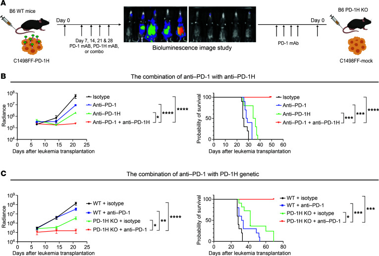
Figure 6. Mouse PD-1H blockade confers a synergistic antileukemic effect with mouse PD-1 blockade.
(A) Syngeneic mouse leukemia model using tail-vein injection with mouse myeloid leukemia cells expressing PD-1H (C1498FF–PD-1H) transplanted into B6 mice, which were then treated with anti–PD-1 and/or anti–PD-1H mAbs. Syngeneic mouse leukemia model using tail-vein injection with mouse myeloid leukemia cells not expressing PD-1H (C1498FF-mock) transplanted into WT B6 mice or PD-1H–KO mice, which were then assessed for in vivo antileukemia effect of genetic deletion of PD-1H in host mice with or without anti–PD-1 mAbs. (B) Synergistic antileukemia effect of anti–PD-1 mAb with anti–PD-1H mAb. In vivo proliferation was assessed by bioluminescence (left) and survival by a Kaplan-Meier plot (right). Radiance indicates the mean value per group, and error bars represent SEM. Data from 2 experiments were combined (n = 10). (C) Synergistic antileukemia effect of genetic deletion of PD-1H in host mice (PD-1H KO) with anti–PD-1 mAb. In vivo proliferation was assessed by bioluminescence (left) and survival by a Kaplan-Meier plot (right). Radiance indicates the mean value per group, and error bars represent SEM. Data from 2 experiments were combined (n = 10). (B and C) P value determined by simple linear regression method for statistical analysis of radiance and log-rank test for survival. *P < 0.05; **P < 0.01; ***P < 0.001; ****P < 0.0001. These experiments were repeated 2 times. Repeated measures were determined by ANOVA with 2 factors (P > 0.05, no difference among experiments).

