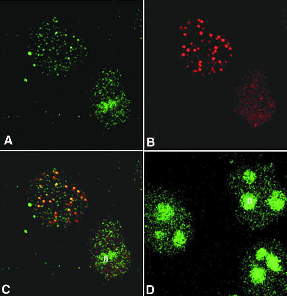
Fig. 4. Co-localization of WRN and RPA proteins in cells arrested in S-phase with HU. (A–C) Cells were arrested in S-phase by HU treatment as described. Following lysis, fixation and permeabilization, cells were incubated with anti-WRN (A; green) or anti-RPA antibodies (B; red). In >50% of the cells, WRN localizes in nuclear foci that coincide with the RPA foci, as illustrated in the superimposed images (C). The yellow colour results from the overlapping of the red and green foci. In cells incubated for the same length of time in the absence of HU, WRN shows the characteristic diffuse nucleolar (n) localization (D).

An official website of the United States government
Here's how you know
Official websites use .gov
A
.gov website belongs to an official
government organization in the United States.
Secure .gov websites use HTTPS
A lock (
) or https:// means you've safely
connected to the .gov website. Share sensitive
information only on official, secure websites.
