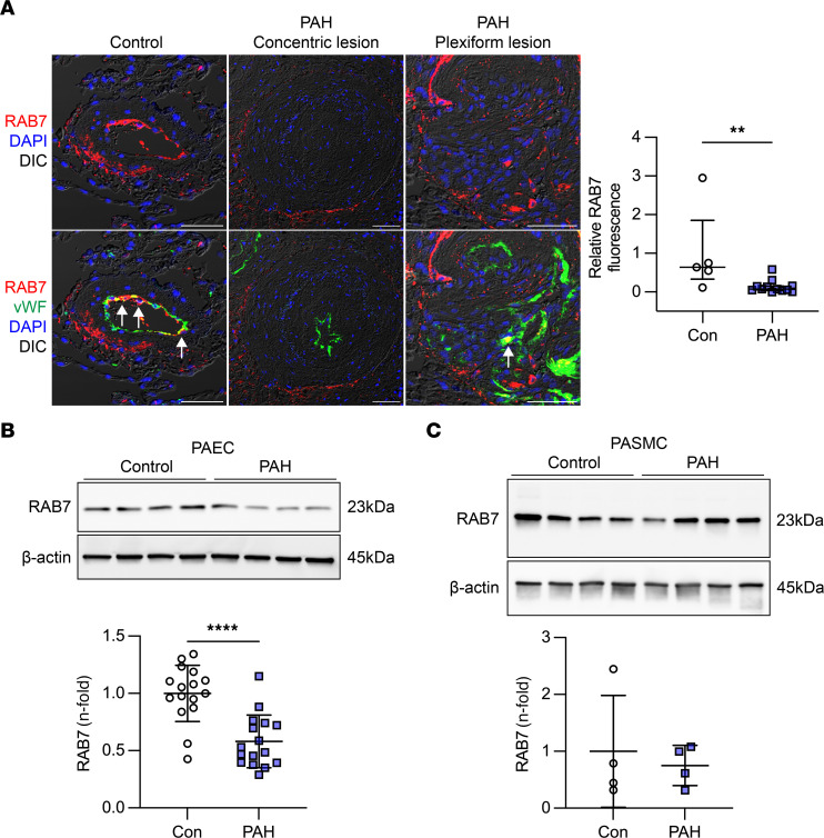
Figure 1. Reduced RAB7 expression in endothelial cells from PAH patients.
(A) Representative optical sections (confocal microscopy, representative of 5 PAs from 3 control [Con] patients and 12 PAs from 3 patients with PAH) show reduced RAB7 expression in ECs in concentric and plexiform lesions from patients with PAH compared with controls. Arrows indicate vWF+ ECs with strong RAB7 expression. Scale bars: 50μm. Nuclei were stained with DAPI. The graph shows quantification of relative RAB7 immunofluorescence in ECs from control and PAH PAs. (B and C) Representative Immunoblot (n = 4 individual control and PAH PAEC lines) and quantification of RAB7 in PAECs (B) in 4 immunoblots from 12 individual controls (failed donors) and 15 individual patients with PAH (n = 16 data points total per group from 4 repeat experiments). (C) Representative immunoblot and quantification of RAB7 in PASMCs from 4 individual controls (failed donors) and 4 individual patients with PAH. β-Actin was used as the loading control. All graphs show single values and the median ± IQR (A) or the mean ± SD (B and C). **P < 0.01 and ****P < 0.0001, by 2-tailed Mann-Whitney U test (A) or 2-tailed Student’s t test (B and C).

