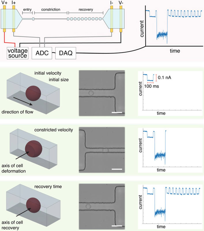
Fig. 5.
Schematic of the operating principles of mechano-node-pore sensing (mechano-NPS), including high-speed micrographs and an example current pulse. First, a cell’s initial size and velocity is measured. The cell then enters a narrower section of the microchannel, where it is constricted along the axis perpendicular to the direction of flow. Finally, the cell is released into a wider pore, where it can recover from its deformed shape to a spherical shape over time. This recovery is reflected by the gradual increase in pulse amplitude. High-speed micrographs show a cell throughout each stage of mechano-NPS; scale bars represent 30 μm

