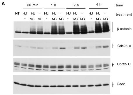
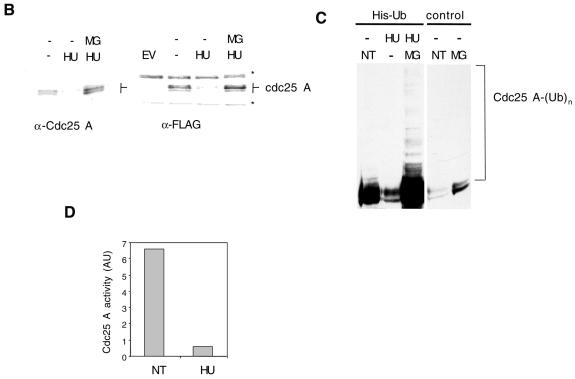
Fig. 2. (A) HeLa cells were treated with 2.5 mM HU, HU plus 5 µM MG132 (HU/MG) or MG132 and lysates (50 µg) analyzed by immunoblotting with the indicated antibodies. NT, untreated cells. (B) HeLa cells (15-cm plates) were transfected with a plasmid encoding Cdc25 A (6 µg) with or without an N-terminal FLAG tag (left and right panel respectively). EV, cells transfected with empty vector. DNA precipitates were left on cells for 15 h. Cells were then split into three plates. After 12 h, the medium was changed and cells were treated with HU or HU plus MG132 for an additional 12 h. Thirty micrograms of lysate were loaded per lane, and blots probed with Cdc25 or FLAG antibodies. Asterisks indicate non-specific bands recognized by the FLAG antibody. (C) Cells were treated as in Figure 1C. Twenty-four hours after removal of the DNA precipitate, cells were either left untreated (NT) or treated for 8 h with 2.5 mM HU or HU plus 5 µM MG132 (MG). (D) Cdc25 A immunoprecipitations were performed on lysates (1 mg) from cells untreated (NT) or treated for 2 h with HU (2.5 mM).

An official website of the United States government
Here's how you know
Official websites use .gov
A
.gov website belongs to an official
government organization in the United States.
Secure .gov websites use HTTPS
A lock (
) or https:// means you've safely
connected to the .gov website. Share sensitive
information only on official, secure websites.

